The imaging of dynamical processes at interfaces and on the nanoscale is of great importance throughout science and technology. While light-optical imaging techniques often cannot provide the necessary spatial resolution, electron-optical techniques damage the specimen and cause dose-induced artifacts. Here, optical near-field electron microscopy (ONEM) is proposed, an imaging technique that combines noninvasive probing with light, with a high-spatial-resolution readout via electron optics. Close to the specimen, the optical near fields are converted into a spatially varying electron flux using a planar photocathode. The electron flux is imaged using low-energy electron microscopy, enabling label-free nanometric resolution without the need to scan a probe across the sample. The specimen is never exposed to damaging electrons.
Raphaël Marchand, Radek Šachl, Martin Kalbáč, Martin Hof, Rudolf Tromp, Mariana Amaro, Sense J. van der Molen, and Thomas Juffmann,
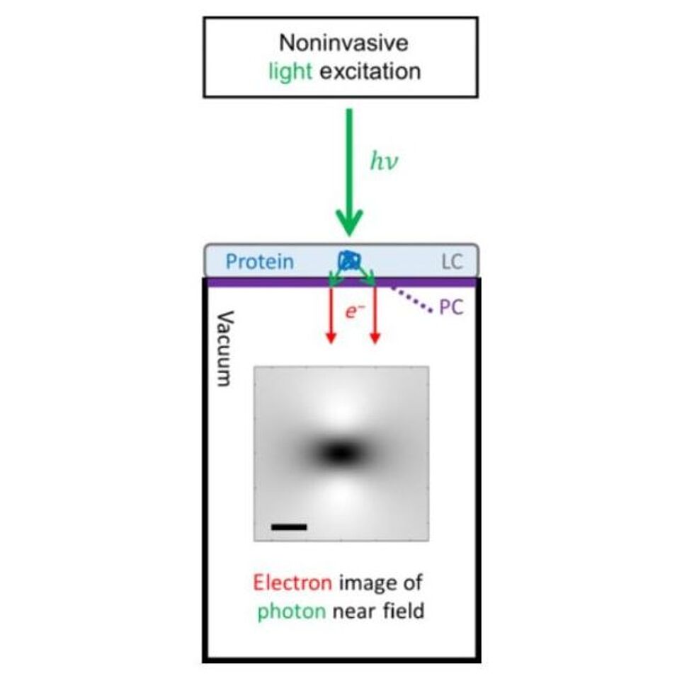
Optical Near-Field Electron Microscopy
Phys. Rev. Applied 16, 014008 (2021).
doi.org/10.1103/PhysRevApplied.16.014008
View full article: here